不同牙周表型成年患者上颌牙列远移前后中切牙区软硬组织的变化
不同牙周表型成年患者上颌牙列远移前后中切牙区软硬组织的变化
作者:陈瑞, 韩爽, 安琪
错畸形是临床上常见的口腔疾病,可影响患者的口颌系统功能,严重者可能会引起牙周病、龋病,对患者的身心健康造成一定危害[1]。正畸医师除了评估牙槽骨重塑的潜力外,还应制定合理的正畸治疗计划,以尽量减少牙周风险[2]。保持牙周健康对于正畸治疗的患者至关重要,牙周组织的稳定是正畸牙齿移动的基础。正畸治疗可以通过消除创伤性咬合和解除拥挤,建立稳定的咬合关系,从而有助于口腔卫生的维护[3],因此,正畸治疗也是改善牙周健康的手段。其中,牙龈表型是需要考虑的一个关键因素,在口腔治疗及预后的结果和可预测性方面起着重要的作用[4]。2017年牙周病世界研讨会上提出:牙周表型包括牙龈表型和骨形态型,牙龈表型指牙龈厚度(gingival thickness,GT)、角化龈宽度(keratinized tissue width,KTW),骨形态型指颊侧骨板厚度(buccal bone plate thickness,BBPT)[5]。依据牙龈厚度通常分为薄龈型和厚龈型2种类型[5- 6]。厚龈型的特征包括角化龈宽,更密集和更多的纤维软组织,大量附着的咀嚼黏膜,骨板厚,骨边缘厚等[7]。研究表明,厚龈型者在应对炎症、机械刺激、治疗的预后等方面相比薄龈型患者有更好的效果[8]。既往研究大多集中在矫治前后牙周硬组织的改建,而忽略了不同牙龈表型的前牙区软硬组织存在差异。因此,本研究从三维方向探讨不同牙龈生物类型在正畸治疗干预下引起的相关牙周组织变化。
1 资料和方法1.1 研究对象
选取2020年9月至2023年9月于合肥市口腔医院正畸科采用非拔牙方法矫治完成的成年错畸形患者52例。依据牙龈厚度分组,厚龈组与薄龈组每组各26例。本研究为回顾性研究,获得合肥市口腔医院伦理委员会的审批(伦理号:YJS20230101)。所有患者均同意将病例资料用于本研究并签署知情同意书。纳入标准:①恒牙列,牙列完整,牙周情况良好,无牙龈红肿、牙齿松动及明显牙槽骨吸收;②安氏Ⅱ类错,前牙覆盖Ⅰ~Ⅱ度;③上牙列轻度拥挤,拥挤度 ≤ 3 mm;④采用微种植支抗钉;⑤无骨代谢性疾病及其他系统性疾病,颞下颌关节检查无明显异常;⑥矫治前后资料完善。排除标准:①服用影响骨代谢的药物;②唇腭裂或唇腭裂治疗史;③妊娠期女性;④服用与牙龈增生风险相关的药物。
1.2 主要实验器材
锥形束计算机断层扫描(cone beam computed tomography,CBCT)(NewTom VGi,Newtom Dental,意大利);头颅侧位摄片机(Planmeca ProMax,Planmeca,芬兰);游标卡尺(MNT-150T,美耐特,德国);牙周探针(PCPUNC156,Hu-Friedy,美国);微种植钉(601-0022,ORMCO,美国)
1.3 研究方法
所有患者在正畸治疗前(T0)、治疗后(T1)分别拍摄CBCT、头颅侧位片,并使用牙周探针测量记录患者双侧上颌中切牙的探诊深度(probing depth,PD)、唇侧角化龈宽度(keratinized tissue width,KTW)等临床指标,单位为mm,游标卡尺测距;牙龈退缩(gingival recession,GR):依据唇侧龈缘中点与釉牙骨质界的位置关系判定,若每例患者有至少1颗上颌中切牙的唇侧龈缘位于釉牙骨质界下方,则有牙龈退缩,记为“1”,若无釉牙骨质界暴露,则无牙龈退缩,记为“0”。
1.3.1 牙周表型的测定及分组
术前依据牙周探针透视法判定牙龈厚度并分组:将牙周探针探入中切牙唇侧中央龈沟内,可视及探针轮廓者为薄龈组,不可视及探针轮廓者为厚龈组。
1.3.2 治疗过程
所有患者在正畸治疗前均进行牙周基础洁治,进行口腔卫生宣教。所有患者若有上颌第三磨牙均需拔除,均采用唇侧矫治系统,每4周更换1次弓丝。使用镍钛丝逐步更换至0.018×0.025英寸不锈钢方丝排齐整平上下牙列。由同1名正畸医师局麻下于患者双侧上颌第一磨牙与第二磨牙之间颊侧膜龈联合处植入长度为10 mm,直径为2 mm的自攻型微种植钉。微种植钉长轴与牙体长轴夹角为45°~70°。所有患者均使用橡皮链(力度:200 g)牵引(图1),每月复诊加力,平均加力时间为9~10个月,远移上牙列直至磨牙、尖牙中性关系,治疗结束后拍摄CBCT及进行上颌中切牙软硬组织变化测量,平均治疗时间为20~22个月。
图1 安氏Ⅱ类错畸形患者上颌全牙列远移示意图
Figure 1 Schematic view of distal migration of maxillary total dentition in patients with Class II malocclusion
1.3.3 CBCT图像测量
对患者使用CBCT机进行头颈部扫描,参数为层厚0.1 mm,扫描时间18~24 s,电压110 kV。将CBCT的DICOM文件导入Mimics21.0软件中进行三维重建,测量治疗前后中切牙软硬组织变化(图2)。
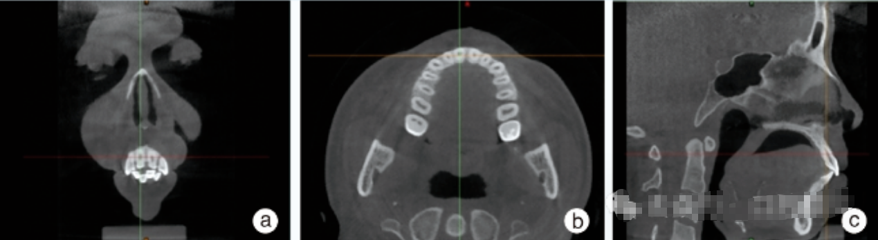
图2 Mimics 21.0软件多平面重建CBCT界面测量治疗前后中切牙软硬组织变化
Figure 2 CBCT measurements of the soft and hard tissue changes of central incisors before and after treatment using Mimics 21.0 software
a: coronal plane; b: transverse plane; c: sagittal plane
Full size|PPT slide
通过调整坐标轴获得矢状面最大径作为相应牙位的测量切面,切缘与根尖点连接得到一条直线,定为牙体长轴(long axis of the tooth, LA)。唇腭侧釉牙骨质界连线(cemento-enamel junction, CEJ)与牙长轴 LA 的交点到根尖点之间的距离定为牙根长度(A),将牙根长度三等分后分别向唇、腭侧做一条垂直于牙长轴的线段,测量该水平唇腭侧牙槽骨厚度,分别记为BW1、BW2、BW3、PW1、PW2、PW3(图3)。测量牙齿唇腭侧牙槽嵴顶沿牙长轴方向至釉牙骨质界距离,即唇腭侧牙槽骨高度,分别记为BH、PH(图3)
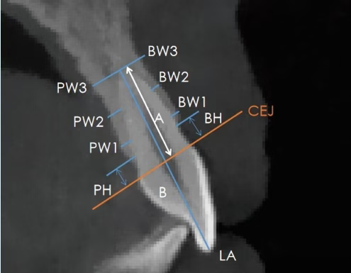
图3 CBCT测量治疗前后唇腭侧牙槽骨厚度、高度示意图
Figure 3 CBCT measurements of labio-palatine alveolar bone thickness and height before and after treatment
LA: long axis of the tooth; A: root length; CEJ: cemento-enamel junction; BW1: labial neck 1/3 alveolar bone thickness; BW2: labial middle 1/3 alveolar bone thickness; BW3: labial tip 1/3 alveolar bone thickness; PW1: lingual neck 1/3 alveolar bone thickness; PW2: lingual middle 1/3 alveolar bone thickness; PW3: lingual tip 1/3 alveolar bone thickness; BH: distance between the crest of the alveolar ridge to cemento-enamel junction; PH: distance from the top to cemento-enamel junction
Full size|PPT slide
1.4 统计学分析
采用 SPSS 26.0 软件对数据进行统计分析,上颌左右中切牙数据差异不具有统计学意义,故本研究将每组研究对象的观测牙数据合并成1组,对合并所得数据进行描述统计。符合正态分布的计量资料采用
均数
标准差表示,组间比较采用独立样本t检验,组内比较采用配对样本t检验。计数资料采用卡方检验进行比较。P < 0.05为差异有统计学意义。
2 结果2.1 一般资料比较
如表1所示,厚龈组中男性13例,女性13例,平均年龄(27.96 ± 4.64)岁;薄龈组中男性9例,女性17例,平均年龄(27.85 ± 4.74)岁。两组患者在年龄、性别方面差异均无统计学意义(P > 0.05)。典型病例见图4、图5。
表1 厚龈组与薄龈组错畸形患者年龄性别比较
Table 1 Comparison of age and sex of malocclusion patients in the thick gingival and thin gingival groups
,n

图4 薄龈组安氏Ⅱ类错畸形患者正畸治疗前后口内照片及锥形束CT
Figure 4 Intraoral images and CBCT of patients with Class II malocclusion in thin gingival group before and after orthodontic treatment
LA: long axis of the tooth; CEJ: cemento-enamel junction; BW1: labial neck 1/3 alveolar bone thickness; BW2: labial middle 1/3 alveolar bone thickness; BW3: labial tip 1/3 alveolar bone thickness; PW1: lingual neck 1/3 alveolar bone thickness; PW2: lingual middle 1/3 alveolar bone thickness; PW3: lingual tip 1/3 alveolar bone thickness; BH: distance between the crest of the alveolar ridge to cemento-enamel junction; PH: distance from the top to cemento-enamel junction; a-c: pretreatment CBCT screenshots and oral photos; female, 22 years old, class II malocclusion, thin gingiva, distal maxillary complete dentition was adopted; d-f: end of treatment CBCT screenshots and oral photos show neutral molar and neutral canine relationships; the thickness of the labial alveolar bone increased in 1/3 and middle 1/3, the apical 1/3 decreased, the palatal alveolar bone decreased in 1/3, the apical 1/3 increased, and the height of alveolar bone decreased the lip and palate side
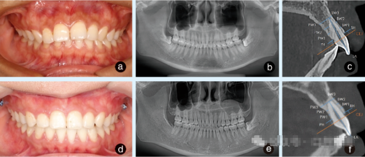
图5 厚龈组安氏Ⅱ类错畸形患者正畸治疗前后口内照片及锥形束CT
Figure 5 Intraoral images and CBCT of patients with Class II malocclusion in the thick gingival group before and after orthodontic treatment
LA: long axis of the tooth; CEJ: cemento-enamel junction; BW1: labial neck 1/3 alveolar bone thickness; BW2: labial middle 1/3 alveolar bone thickness; BW3: labial tip 1/3 alveolar bone thickness; PW1: lingual neck 1/3 alveolar bone thickness; PW2: lingual middle 1/3 alveolar bone thickness; PW3: lingual tip 1/3 alveolar bone thickness; BH: distance between the crest of the alveolar ridge to cemento-enamel junction; PH: distance from the top to cemento-enamel junction; a-c: pretreatment CBCT screenshots and oral photos; female, 25 years old, class II malocclusion, thick gingiva, distal maxillary complete dentition was adopted; d-f: end of treatment CBCT screenshots and oral photos show neutral molar and neutral canine relationships; the thickness of the labial alveolar bone increased in 1/3 and middle 1/3, the apical 1/3 decreased, the palatal alveolar bone decreased in 1/3, and the apical 1/3 increased, and the height of alveolar bone decreased the lip and palate side
2.2 治疗前后硬组织X线头影测量结果
组内比较显示,正畸治疗后厚龈组SNA、SNB、ANB减小,但差异无统计学意义(P > 0.05);U1-NA减小,差异有统计学意义(P < 0.05)。薄龈组SNA、SNB、ANB减小,但差异无统计学意义(P > 0.05);U1-NA减小,差异有统计学意义(P < 0.05),见表2。
表2 厚龈组与薄龈组错畸形患者治疗前后硬组织X线片头影测量结果
Table 2 Results of hard tissue X-ray head shadow measurements before and after treatment in patients in the thick gingival and thin gingival groups
, n=26

T0: initial situation; Tl: final situation; S: sella; N: nasion; A: subspinale; B: supramental; U1: upper incisor; NA: the plane of N-A; SNA: sella-nasion-subspinale angle; SNB: sella-nasion-supramental angle; ANB: subspinale-nasion-supramental angle; U1-NA/°: the angle between the long axis of the upper central incisor and the NA line; U1-NA/mm: the vertical distance between the incisor edge of the upper central incisor and the NA line
2.3 两组治疗前后硬组织X线头影测量数据变化量对比
组间比较显示,治疗后两组间SNA、SNB、ANB、U1-NA变化量差异均无统计学意义(P > 0.05),见表3。
表3 厚龈组与薄龈组错畸形患者治疗前后硬组织X线头影测量结果变化量的对比
Table 3 Comparison of the change of hard tissue X-ray head shadow measurements before and after treatment in patients in the thick gingival and thin gingival groups
, n=26
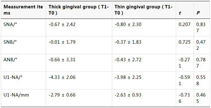
T0: initial situation; Tl: final situation; S: sella; N: nasion; A: subspinale; B: supramental; U1: upper incisor; NA: the plane of N-A; SNA: sella-nasion-subspinale angle; SNB: sella-nasion-supramental angle; ANB: subspinale-nasion-supramental angle; U1-NA/°: the angle between the long axis of the upper central incisor and the NA line; U1-NA/mm: the vertical distance between the incisor edge of the upper central incisor and the NA line
2.4 治疗前后上颌中切牙唇腭侧牙槽骨厚度和高度变化
如表4所示,正畸治疗后,两组唇侧牙槽骨在颈1/3、中1/3处均增厚,差异有统计学意义(P < 0.05),而在根尖1/3处牙槽骨厚度下降,差异有统计学意义(P < 0.05);两组腭侧牙槽骨在颈1/3、中1/3处均变薄,差异有统计学意义(P < 0.01),在根尖1/3处牙槽骨厚度增加,差异有统计学意义(P < 0.01);两组唇腭侧牙槽嵴顶到CEJ的距离均增加,表示唇腭侧牙槽骨高度均降低,其中唇侧牙槽骨高度下降较少,差异无统计学意义(P > 0.05),而腭侧牙槽骨高度则降低明显,差异有统计学意义(P < 0.05)。
表4 厚龈组与薄龈组错畸形患者治疗前后上颌中切牙唇腭侧牙槽骨厚度和高度变化
Table 4 Alveolar bone thickness and height of maxillary central incisor lip and palate of the thick gingival and thin gingival groups
, n=26

T0: initial situation; Tl: final situation; BW1: labial neck 1/3 alveolar bone thickness; BW2: labial middle 1/3 alveolar bone thickness; BW3: labial tip 1/3 alveolar bone thickness; PW1: lingual neck 1/3 alveolar bone thickness; PW2: lingual middle 1/3 alveolar bone thickness; PW3: lingual tip 1/3 alveolar bone thickness; BH: distance between the crest of the alveolar ridge to cemento-enamel junction; PH: distance from the top to cemento-enamel junction
2.5 两组治疗前后上颌中切牙牙槽骨变化量对比
如表5所示,组间比较显示,薄龈组腭侧牙槽骨高度的下降显著,差异有统计学意义(P < 0.05),其他差异均无统计学意义(P > 0.05)。
表5 厚龈组与薄龈组错畸形患者治疗前后上颌中切牙牙槽骨变化量对比
Table 5 Comparison of changes of alveolar bone maxillary central incisor before and after treatment between patients in the thick gingival and thin gingival groups
, n=26
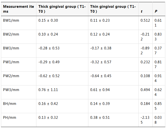
T0: initial situation; Tl: final situation; BW1: labial neck 1/3 alveolar bone thickness; BW2: labial middle 1/3 alveolar bone thickness; BW3: labial tip 1/3 alveolar bone thickness; PW1: lingual neck 1/3 alveolar bone thickness; PW2: lingual middle 1/3 alveolar bone thickness; PW3: lingual tip 1/3 alveolar bone thickness; BH: distance between the crest of the alveolar ridge to cemento-enamel junction; PH: distance from the top to cemento-enamel junction
2.6 治疗前后上颌中切牙牙周参数变化
如表6所示,正畸治疗后,两组上颌中切牙PD、KTW、GR差异均无统计学意义(P > 0.05)。
表6 厚龈组与薄龈组错畸形患者正畸治疗前后上颌中切牙牙周软组织变化
Table 6 Changes of periodontal soft tissue of maxillary central incisor before and after orthodontic treatment in patients in the thick gingival and thin gingival groups
, n=26

T0: initial situation; Tl: final situation; PD: probing depth; GR:gingival recession; KTW: keratinized tissue width
2.7 两组治疗前后上颌中切牙牙周参数变化量对比
如表7 所示,正畸治疗后,两组上中切牙PD、KTW、GR变化量差异均无统计学意义(P > 0.05)。
表7 厚龈组与薄龈组错畸形患者治疗前后上颌中切牙牙周参数变化量对比
Table 7 Comparison of changes in the periodontal parameters of maxillary central incisor before and after treatment between patients in the thick gingival and thin gingival groups
, n=26
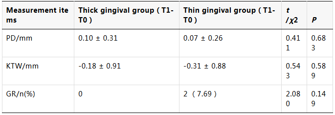
T0: initial situation; Tl: final situation; PD: probing depth; GR: gingival recession; KTW: keratinized tissue width
3 讨论
本研究使用探针透视法根据牙龈厚度不同将受试者分为薄龈、厚龈两组,临床上常用此法对牙龈表型进行分型,此种方法可重复性好、无创,临床上易于操作[9]。目前牙龈表型与性别之间的相关性尚不明确,Vandana等[10]认为下颌前牙区的唇侧牙龈厚度男性厚于女性,但在上颌前牙区无统计学差异,这与本研究结果相一致。
传统2D射线照相术具有影像重叠、精度差、不能提供三维图像的缺点,这使得口腔医师难以准确评估解剖结构的尺寸和位置关系,影响诊断和治疗计划的制定[11]。此外,了解骨密度对于评估牙齿移动的可行性和牙槽骨的潜力至关重要[12],而2D射线照相术提供的二维图像可能无法准确反映骨的真实密度和体积。气道评估也是正畸诊断和治疗计划的一个重要组成部分,然而2D图像在准确评估气道的大小和形态方面也存在一定局限性[13]。随着正畸实践的不断发展,CBCT已成为一项强大的工具,被广泛用于扫描颅面组织[14]。因此,本研究通过采用CBCT结合逆向工程软件Mimics Research 21.0软件对牙槽骨的三维变化进行定量测量,大大提高了测量的精确性。
正畸牙齿移动范围受牙槽骨板厚度的影响,薄龈型患者牙槽骨板相对较薄,牙齿移动若超出牙槽骨板可能导致骨开窗、骨开裂及牙槽骨垂直方向上的丧失[15]。本研究结果表明,薄龈组术前牙槽骨厚度较薄,尤其是唇侧牙颈部,这与以往的研究结果相一致[16],提示在正畸治疗过程中,薄龈者出现牙周问题的风险更大。有研究表明,上前牙在内收移动过程中,唇腭侧牙槽骨总厚度和切牙区牙槽骨面积减少,即牙齿在移动过程中发生“穿骨移动”[17]。本研究结果显示,上牙列整体远移后,牙槽骨的改建并不均匀,两组上中切牙牙槽骨均表现为在唇侧颈1/3、中1/3处以及腭侧根尖1/3处厚度增加,而在唇侧根尖1/3处,腭侧颈1/3以及根中1/3处厚度减少。这提示在上牙列远移过程中,即使加大了对切牙的转矩控制,也难以使牙齿实现整体移动,而是有控制的倾斜移动。究其原因,尽管本研究采用微种植钉结合牵引钩内收上切牙,但受制于上颌颧牙槽嵴处膜龈联合位置及微种植钉植入高度等因素,在内收前牙时,内收力的作用线仍位于上颌中切牙阻抗中心的方。上切牙在远移过程中发生倾斜移动时,牙周组织内的应力分布并不均匀,唇侧的正畸应力集中于近根尖区,腭侧的应力则集中于近牙槽嵴顶区,致使上切牙唇腭侧牙槽骨的改建并不一致。许多学者的研究表明,牙齿移动后,其周围牙槽骨的重塑与吸收并不完全同步[18-19]。李鹏等[20]认为骨皮质和骨松质的改建速率不一致,前牙内收后,唇侧靠近牙根表面的松质骨增生活跃,而靠近外表面的皮质骨吸收缓慢,最终表现为唇侧骨厚度总体增加。本研究结果进一步支持了这一结论,而成人的风险更高,因为其骨膜中成骨细胞的数量和再生能力随着年龄的增长而降低[21],因此,临床医生在制定治疗计划时,不应设计过大距离的牙齿运动,以避免将牙根置于牙槽骨边界之外。此外,正畸治疗后两组上颌中切牙牙槽骨高度在唇腭侧均有不同程度的降低,其中薄龈组腭侧牙槽骨高度降低量更为显著,这与Kim等[22]的结果相似,上前牙在移动过程中骨增量小于骨吸收,腭侧牙槽骨表现以吸收为主。既往的研究表明,正畸治疗后牙槽骨高度降低,骨开裂的频率增加,与正畸力的施力方向具有一定相关性[23]。其他研究同样发现,薄龈生物型者在前牙大量内收后,腭侧牙槽骨损失更明显,这提示薄龈者在正畸治疗后上切牙腭侧发生骨开裂、骨开窗的风险更高[24]。分析其原因可能是薄龈生物型者骨板较薄,前牙在内收过程中,腭侧牙槽骨受力吸收后,使阻力中心更接近牙尖方向,牙齿倾斜角度增大,但这需要更进一步的实验验证。总之,正畸医生在制定正畸治疗计划时,不仅需要考虑牙齿移动方式以及牙周组织潜力等因素,更不可忽视牙周表型的重要性。
牙龈退缩在临床上一直受到正畸医生的广泛关注。不恰当的正畸治疗作为一种医源性因素,其与牙龈退缩之间的联系已被反复提出[25-26]。Fleming等[27]研究认为正畸治疗后,牙龈厚度是发生牙龈退缩的重要相关因素。Koppolu等[28],Alkan等[29]发现,牙龈退缩的发生与牙龈厚度、角化龈宽度之间存在负相关,即牙龈厚度越厚,角化龈宽度越大,发生牙龈退缩的风险越低。Das等[30]也认为,牙龈退缩与组织生物型或基底骨板厚度密切相关,当薄龈生物型者接受正畸治疗后,更容易发生角化龈宽度丧失、牙龈退缩。但本研究结果显示,两组上切牙在正畸治疗后,PD、KTW、GR差异均无统计学意义。此外,本研究结果表明在上牙列远移过程中上中切牙唇倾度得到了较好的改善。有研究发现,正畸牙唇侧的牙龈退缩更可能是由于牙齿发生了唇倾而不是舌倾引起的,牙齿的倾斜度每增加1°,牙龈退缩将增加约0.2 mm,并且牙齿的倾斜移动会使牙根接近于皮质骨,这可能导致骨变薄并增加骨开窗、骨开裂的风险[31-32]。
正畸治疗后牙周组织改建需要一定的时间进行保持及稳定,而牙槽骨在保持期间的修复能力目前尚存在争议。Sarikaya等[33]发现,在保持阶段的4个月后牙槽骨发生骨沉积,但没有恢复到治疗前的水平。相反,Ahn等[34]在保持期间未观察到新形成的骨。这也是本研究存在的局限性,未来将增加对治疗后患者的长期随访。
综上所述,上颌中切牙在内收过程中唇腭侧牙槽骨改建并不均匀,腭侧牙槽骨以吸收为主,临床上需加以重视;牙周表型是影响正畸治疗后牙周组织改建的重要因素,薄龈型者正畸治疗后腭侧牙槽骨高度降低更明显,发生骨开窗、骨开裂的风险更大。
【Author contributions】 Chen R conceived and authored the article. Han S and An Q contributed to the conception and review of the article. All authors read and approved the final manuscript as submitted.
参考文献
[1]
Rafiq T, Aqeel R, Javed M, et al. Diet and oral health: an investigation into the impact of pakistani dietary habits on oral disease prevalence[J]. Cureus, 2024, 16(10): e71423. doi.org/10.7759/cureus.71423.
本文引用 [1]
[2]
Mao H, Yang A, Pan Y, et al. Displacement in root apex and changes in incisor inclination affect alveolar bone remodeling in adult bimaxillary protrusion patients: a retrospective study[J]. Head Face Med, 2020, 16(1): 29. doi:.10.1186/s13005-020-00242-2.
https://doi.org/10.1186/s13005-020-00242-2
https://www.ncbi.nlm.nih.gov/pubmed/33213460
本文引用 [1] 摘要
[3]
Alasiri MM, Almalki A, Alotaibi S, et al. Association between gingival phenotype and periodontal disease severity-a comparative longitudinal study among patients undergoing fixed orthodontic therapy and invisalign treatment[J]. Healthcare(Basel), 2024, 12(6): 656.doi: 10.3390/healthcare12060656.
本文引用 [1]
[4]
Herrera-Serna BY, López-Soto OP, Chacón T, et al. Relationship between the gingival biotype and the results of root covering surgical procedures: a systematic review[J]. J Clin Exp Dent, 2022, 14(9): e762-e768. doi: 10.4317/jced.59783.
本文引用 [1]
[5]
Jepsen S, Caton J G, Albandar J M, et al. Periodontal manifestations of systemic diseases and developmental and acquired conditions: consensus report of workgroup 3 of the 2017 world workshop on the classification of periodontal and peri‐implant diseases and conditions[J]. J Clin Periodontol, 2018, 45 (Suppl) 20:S219-S229.doi: 10.1111/jcpe.12951.
本文引用 [2]
[6]
Malpartida-Carrillo V, Tinedo-Lopez PL, Guerrero ME, et al. Periodontal phenotype: a review of historical and current classifications evaluating different methods and characteristics[J]. J Esthet Restor Dent, 2021, 33(3): 432-445. doi: 10.1111/jerd.12661.
本文引用 [1]
[7]
Abdelhafez RS, Mustafa NM. Determining the periodontal phenotype-probe transparency versus actual: a diagnostic study[J]. J Esthet Restor Dent, 2023, 35(7): 1001-1007. doi: 10.1111/jerd.13032.
https://www.ncbi.nlm.nih.gov/pubmed/36891868
本文引用 [1] 摘要
[8]
Kim DM, Bassir SH, Nguyen TT. Effect of gingival phenotype on the maintenance of periodontal health: an american academy of periodontology best evidence review[J]. J Periodontol, 2020, 91(3): 311-338. doi: 10.1002/JPER.19-0337.
https://www.ncbi.nlm.nih.gov/pubmed/31691970
本文引用 [1] 摘要
[9]
Aslan S, Clauser T, Testori T, et al. A novel technique for the estimation of gingival thickness: a preliminary study[J]. Int J of Periodontics & Restorative Dent, 2021, 41(4): 571-577. doi: 10.11607/prd.4947.
本文引用 [1]
[10]
Vandana KL, Savitha B. Thickness of gingiva in association with age, gender and dental arch location[J]. J Clin Periodontol, 2005, 32(7): 828-830. doi: 10.1111/j.1600-051X.2005.00757.x.
https://www.ncbi.nlm.nih.gov/pubmed/15966893
本文引用 [1] 摘要
[11]
Polizzi A, Serra S, Leonardi R. Use of CBCT in orthodontics: a scoping review[J]. J Clin Med, 2024, 13(22): 6941.doi: 10.3390/jcm13226941.
本文引用 [1]
[12]
Lo Giudice A, Leonardi R, Ronsivalle V, et al. Evaluation of pulp cavity/chamber changes after tooth-borne and bone-borne rapid maxillary expansions: a CBCT study using surface-based superimposition and deviation analysis[J]. Clin Oral Investig, 2021, 25(4): 2237-2247. doi: 10.1007/s00784-020-03539-3.
本文引用 [1]
[13]
Lo Giudice A, Ronsivalle V, Gastaldi G, et al. Assessment of the accuracy of imaging software for 3D rendering of the upper airway, usable in orthodontic and craniofacial clinical settings[J]. Prog Orthod, 2022, 23(1): 22. doi: 10.1186/s40510-022-00413-8.
https://www.ncbi.nlm.nih.gov/pubmed/35691961
本文引用 [1] 摘要
[14]
Sehrawat S, Sidhu MS, Grover S, et al. “A century of orthodontic progress”-innovations in orthodontics[J]. Orthod J Nepal, 2021, 11(1): 65-71. doi: 10.3126/ojn.v11i1.39091.
本文引用 [1]
[15]
Yildiz MS, Ulutas PA, Ozenci I, et al. Clinical and radiographic assessment of the association between orthodontic mini-screws and periodontal health[J]. BMC Oral Health, 2024, 24(1): 1376. doi: 10.1186/s12903-024-05136-2.
本文引用 [1]
[16]
Ashfaq M, Sadiq A, Sukhia RH, et al. Association of hard and soft tissue factors with gingival recession in orthodontically treated patients: a retrospective study[J]. Int Orthod, 2021, 19(1): 60-66.doi: 10.1016/j.ortho.2020.12.001.
本文引用 [1]
[17]
Zheng Y, Zhu C, Zhu M, et al. Difference in the alveolar bone remodeling between the adolescents and adults during upper incisor retraction: a retrospective study[J]. Sci Rep, 2022, 12(1): 9161. doi:10.1038/s41598-022-12967-y.
https://www.ncbi.nlm.nih.gov/pubmed/35650260
本文引用 [1] 摘要
[18]
Hung BQ, Hong M, Kyung HM, et al. Alveolar bone thickness and height changes following incisor retraction treatment with microimplants[J]. Angle Orthod, 2022, 92(4): 497-504.doi: 10.2319/091121-702.1.
https://www.ncbi.nlm.nih.gov/pubmed/35230383
本文引用 [1] 摘要
[19]
Elnagar MH, Handelman CS, Lippincott JS, et al. Alveolar cortical plate changes associated with incisor retraction and its influence on the limits of orthodontic tooth movement[J]. Orthod Craniofac Res, 2021, 24(4): 536-542.doi: 10.1111/ocr.12469.
https://www.ncbi.nlm.nih.gov/pubmed/33475228
本文引用 [1] 摘要
[20]
李鹏, 孙建伟, 白煜, 等. 青少年内收上切牙后牙槽骨改建的研究[J]. 实用口腔医学杂志, 2019, 35(2): 239-242. doi: 10.3969/j.issn.1001-3733.2019.02.016.
Li P, Sun JW, Bai Y, et al. The remodeling of alveolar bone due to retraction of upper incisors in adolescents[J]. J Pract Stomatol, 2019, 35(02): 239-242. doi: 10.3969/j.issn.1001-3733.2019.02.016.
本文引用 [1]
[21]
Ferretti C, Lucarini G, Andreoni C, et al. Human periosteal derived stem cell potential: the impact of age[J]. Stem Cell Rev Rep, 2015, 11(3): 487-500. doi: 10.1007/s12015-014-9559-3.
https://www.ncbi.nlm.nih.gov/pubmed/25239138
本文引用 [1] 摘要
[22]
Kim HJ, Noh HK, Park HS. Recovery bone formation on radiographic palatal bone dehiscences after incisor retraction with microimplants[J]. Angle Orthod, 2024, 94(2): 168-179. doi: 10.2319/081823-566.1
本文引用 [1]
[23]
Castro LO, Castro IO, de Alencar AH, et al. Cone beam computed tomography evaluation of distance from cementoenamel junction to alveolar crest before and after nonextraction orthodontic treatment[J]. Angle Orthod, 2016, 86(4): 543-549.doi: 10.2319/040815-235.1.
https://www.ncbi.nlm.nih.gov/pubmed/26379114
本文引用 [1] 摘要
[24]
Chanmanee P, Charoemratrote C. Maxillary anterior gingiva and dentoalveolar changes after en-masse retraction between thick and thin gingival biotypes[J]. Am J Orthod Dentofacial Orthop, 2022, 161(6): 838-848. doi: 10.1016/j.ajodo.2021.01.036.
本文引用 [1]
[25]
Niemczyk W, Niemczyk S, Prokurat M, et al. Etiology of gingival recession-a literature review[J]. Wiad. Lek, 2024, 77(5): 1080-1085.doi: 10.36740/WLek202405131.
本文引用 [1]
[26]
Luchian I, Surlari Z, Goriuc A, et al. The influence of orthodontic treatment on periodontal health between challenge and synergy: a narrative review[J]. Dent J(Basel), 2024, 12(4): 112.doi: 10.3390/dj12040112.
本文引用 [1]
[27]
Fleming PS, Andrews J. The role of orthodontics in the prevention and management of gingival recession[J]. Br Dent J, 2024, 237(5): 341-347. doi: 10.1038/s41415-024-7781-1.
本文引用 [1]
[28]
Koppolu P, Al Arabi AA, Al Khayri MJ, et al. Correlation between gingival thickness and occurrence of gingival recession[J]. J Pharm Bioallied Sci, 2023, 15(Suppl 1): S495-S501. doi: 10.4103/jpbs.jpbs_585_22.
https://www.ncbi.nlm.nih.gov/pubmed/37654274
本文引用 [1] 摘要
[29]
Alkan Ö, Kaya Y, Tunca M, et al. Changes in the gingival thickness and keratinized gingival width of maxillary and mandibular anterior teeth after orthodontic treatment[J]. Angle Orthod, 2021, 91(4): 459-467. doi: 10.2319/092620-820.1.
https://www.ncbi.nlm.nih.gov/pubmed/33625489
本文引用 [1] 摘要
[30]
Das G, Ahmed AR, Suleman G, et al. A comparative evaluation of dentogingival tissue using transgingival probing and cone-beam computed tomography[J]. Medicina(Kaunas), 2022, 58(9): 1312.doi: 10.3390/medicina58091312.
本文引用 [1]
[31]
Alsulaimani L, Qali M. Relationship between cone-beam CT evaluation and vlinical evaluation before and after orthodontic treatment and the rate of gingival recession: a systematic review[J]. Cureus, 2024, 16(6):e62536. doi: 10.7759/cureus.62536.
本文引用 [1]
[32]
谢飘, 唐镇, 刘剑. 拔牙矫治后前牙区牙槽骨变化的研究进展[J]. 口腔疾病防治, 2023, 31(6): 452-456. doi: 10.12016/j.issn.2096-1456.2023.06.011.
摘要
Xie P, Tang Z, Liu J. Research progress on anterior alveolar bone changes after orthodontic treatment involving extractions[J]. J Prev Treat Stomatol Dis, 2023, 31(6): 452-456. doi: 10.12016/j.issn.2096-1456.2023.06.011.
本文引用 [1] 摘要
[33]
Sarikaya S, Haydar B, Ciǧer S, et al. Changes in alveolar bone thickness due to retraction of anterior teeth[J]. Am J Orthod Dentofacial Orthop, 2002, 122(1): 15-26. doi: 10.1067/mod.2002.119804.
本文引用 [1]
[34]
Ahn HW, Moon SC, Baek SH. Morphometric evaluation of changes in the alveolar bone and roots of the maxillary anterior teeth before and after en masse retraction using cone-beam computed tomography[J]. Angle Orthod, 2013, 83(2): 212-221.doi: 10.2319/041812-325.1.
本文引用 [1]

 邓超牙齿是烤瓷牙
 娱乐圈整过牙的明
 一群曾经牙齿丑得