
阻生第三磨牙拔除过程中的意外移位 智齿掉进间隙
阻生第三磨牙拔除过程中的意外移位 智齿掉进间隙
门诊手术干预治疗阻生第三磨牙可能伴随多种潜在并发症,发生率为4.6%至30.9%[1,2]。阻生第三磨牙拔除过程中的意外移位已被广泛认知,但关于其发生率及处理的文献相对匮乏。尽管属于罕见事件,但其可能导致严重甚至危及生命的并发症。舌向位阻生第三磨牙常伴随舌侧骨板的开窗性缺损,若使用牙挺时施力不当,易导致牙齿意外移位至原发或继发间隙。
Thoma 提出,根尖周感染导致的舌侧骨板菲薄易引发穿孔 [3]。Stacy描述了下颌舌侧骨板自然穿孔的发生率,认为其是牙体或牙根移位的易感因素 [4]。
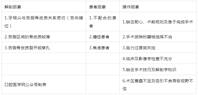
Chubb DWR等指出,患者相关解剖因素及术者操作因素可能促使牙体/牙根碎片移位至下颌下间隙 [5](表1)。
表1
影响牙齿移位的因素
下颌阻生磨牙最常见的移位部位为下颌下间隙及翼下颌间隙[6]。由于操作空间有限、视野不佳且存在出血风险,移位牙的手术处理过程可能较为复杂。
本文旨在报道1例下颌第三磨牙移位至下颌下间隙的病例,并回顾相关文献,以制定在基层医疗环境中的处理指南。
病例报告
患者,女,33岁,就诊于Santosh牙科学院口腔颌面外科,主诉左下后牙区持续疼痛7天。既往体健,无全身系统性疾病。口外检查示左侧下颌下淋巴结触痛且肿大。口内检查可见左下第三磨牙存在明显龋坏。详细影像学评估显示,该牙为远中倾斜、A 位、II 类阻生第三磨牙,Pederson难度指数评分为7,提示手术“极具难度”(图1a)。下牙槽神经管白线连续性中断,提示与下牙槽神经“邻近”。治疗方案拟沿釉牙骨质界从冠部截冠以开辟脱位通道,再分根拔除。已向患者解释手术的利弊及潜在并发症,并获得患者的书面知情同意。

图1
a 术前全景片(OPG)显示远中倾斜的 38 牙严重龋坏
b 掀起 Widman 瓣后行颊侧去骨
c 移位的牙根碎片位于左下颌下间隙近下颌骨下缘处
d 牙根碎片位于下颌舌骨肌深面
e 取出的牙根碎片
f 术后全景片
g 制备的 CGF 凝胶填充拔牙窝
使用碘伏进行口内外消毒准备,患者以无菌方式铺巾。采用2%利多卡因肾上腺素注射液行舌神经、颊长神经及下牙槽神经阻滞麻醉。
使用15号 BP 刀片及3号 BP 刀柄,自第二磨牙远颊侧垂直松弛切口,沿阻生牙牙龈缘作龈沟切口,远中附加松弛切口,翻起梯形粘骨膜瓣。暴露阻生牙后,使用标准外科微电机手机及外科车针(SS White HP 702)在大量生理盐水冲洗下进行颊侧去骨(图1b)。
尝试挺出牙体时发生牙冠部断裂,残冠被取出。多次尝试用牙挺取出断根未果,仅取出少量牙根碎片及周围骨组织。随后发现拔牙窝内牙根碎片不慎丢失且无法定位(图2B)。
对拔牙窝进行清创和冲洗时,观察到舌侧骨皮质穿孔。通过示指触诊舌侧确认连续性中断,但未触及牙根碎片。拍摄检查 X 线片(全景片)定位缺失碎片,显示下颌骨下缘存在放射阻射影,提示牙根碎片移位至下颌角邻近的下颌下间隙内(图1c)。
向患者说明并发症及取出断根的必要性后,决定局麻下即刻取出断根。

图2
A 下颌第三磨牙区域解剖示意图
B 智齿牙根移位至下颌下间隙
C 取出移位的牙根碎片
(a. 舌神经;b. 下牙槽神经;c. 下颌舌骨肌;d. 穿孔 / 骨折的舌侧骨皮质;e. 移位的牙根碎片;f. 使用 Howarth 剥离器暴露并稳定碎片)
计划行宽基底舌侧瓣。自侧切牙至第三磨牙区沿舌侧龈缘作沟内切口,在舌侧翻起全厚信封瓣。小心剥离下颌舌骨肌附着以暴露下颌下间隙。在直视下可触及断根位于下颌下缘邻近区(图1d)。以豪沃氏骨膜分离器稳定断根防止其向深部移位(图2C),后以蚊式钳取出(图1e)。术后OPG确认断根完全取出(图1f)。
手术部位用碘伏和生理盐水冲洗后,使用 SS White 圆车针(HP 6号)修整锐利骨缘。随后将术前采集的10 ml 血液用于制备浓缩生长因子(CGF),填充拔牙窝以加速愈合过程(图1g)。使用3-0丝线间断缝合黏膜瓣实现一期闭合。开具5天疗程的抗生素和镇痛药,术后第7天拆线。术后第10天,患者无不适,恢复良好。
讨论
与其他手术一样,智齿的手术拔除可能存在风险或并发症。这些操作常伴随术后疼痛、肿胀和张口受限等暂时性并发症[7]。
文献显示,出血、干槽症、感染和暂时性感觉异常是最常见的副作用。尽管罕见,但牙槽突骨折、第三磨牙移位至筋膜间隙、神经损伤(下牙槽神经/舌神经)[8]、口腔上颌窦交通或下颌骨骨折等并发症仍对患者构成风险,亦增加术者操作难度 [9–12]。
本文建议采用以下流程处理颌下间隙移位牙/根:
- 彻底清创拔牙窝以改善视野,避免盲目操作导致断端向深部移位;
- 指诊确认舌侧骨板连续性中断时,应怀疑断端移入舌下、下颌下或翼下颌间隙;
- 需在两个平面拍摄OPG及咬合片进行定位,若无法定位则推荐CT/CBCT [13];
- 定位成功后应经患者同意安排手术(图3)。
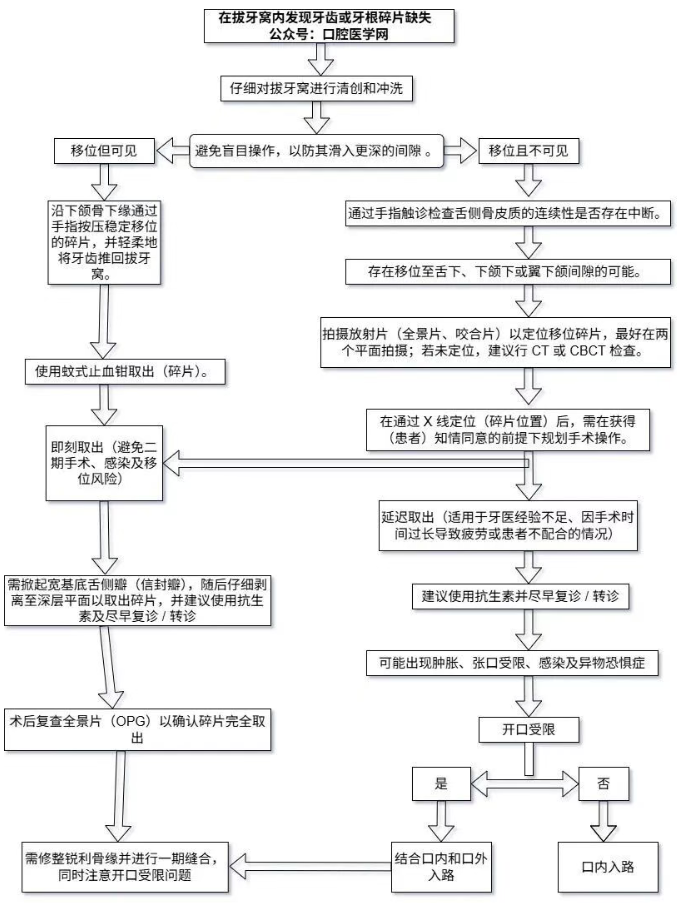
图3
拔牙窝中碎片缺失的处理流程
文献中记载了多种取出技术。
常规术式:通过下颌升支至前磨牙区翻起舌侧粘骨膜瓣,经口内入路取出。局麻下此方法创伤最小且操作简便,但可能暴露不足;
联合入路:当视野受限、舌神经损伤及出血风险较高时,可采用口内外联合入路处理深部移位。优势在于操作成功率较高,但需口外切口及遗留瘢痕;
导航技术:计算机辅助内镜导航技术安全高效且微创,但设备昂贵且普及受限 [14–16]。
1958年Howe首次报道牙齿意外移位病例,提出口底移位牙取出方法 [17](表2,略)。此后零星报道多例第三磨牙移位及处理方案。
2002年Yeh等提出口内外联合术式:口内翻宽基舌侧瓣联合下颌下区4mm皮肤切口,以止血钳及凯利钳取出断端,3例均无并发症 [30];
2007年Huang等综述25篇文献(1958–2005),提出改良术式:拔牙窝舌侧骨板截骨联合软组织剥离以改善视野 [46];
2014年Jolly等强调局麻下口内处理下颌下间隙移位牙根前需全面评估风险因素 [35];
2016年Solanki报道2例下颌下间隙移位牙取出术,其中1例并发舌神经感觉异常(3个月恢复)[38];Adeyemi报道1例移位牙滞留1月后继发咬肌下脓肿 [37];
2019年Di Nardo等综述68篇文献:19例移位至颞下窝,11例至舌下间隙,9例至下颌下间隙,11例至咽旁间隙,并分析各间隙解剖结构、诱发因素及并发症 [47];
2021年Chattopadhyay等强调早期干预可降低发病率 [42];Chubb等提出舌侧瓣设计及翻瓣时保护舌神经的要点,并制定下颌下间隙移位牙处理流程 [5];
2023年Nadeem等指出拔牙时过度施力是移位的主因 [43];Konate等提出下颌下间隙移位牙处理的决策树 [45]。
文献统计(1958–2023)33篇报道共44例下颌下间隙牙体/牙根移位(牙冠碎片25例,牙根19例)。仅3篇报道即刻取出,其余均为延迟处理(多因基层诊所转诊)。
本机构病例中,操作急躁及暴力挺出是移位主因。建议识别风险因素后谨慎操作,正如Howe所言:"急躁是口腔外科之敌"。
本病例断根移位后选择即刻经口内宽基舌侧瓣入路,视野暴露充分 [16]。推荐断端移位至下颌下间隙后立即取出,以降低感染/纤维化风险,避免患者因残留断端产生恐惧心理。
关于取出时机存在一些争议,部分学者认为延迟干预可能有利于碎片纤维化和稳定,而另一些学者则认为延迟取出可能导致疼痛、肿胀、张口受限、心理困扰、舌神经感觉异常、感染和移行等并发症。
基层条件下建议:
-OPG为首选定位手段[48] ;
根尖片(胶片/RVG传感器)需要放置在舌侧沟更深的位置,可能导致进一步移位 [49];
全景片联合咬合片可双平面定位;
后前位及颏顶位片 [12] 可作为补充;
下颌下间隙移位首选舌侧信封粘骨膜瓣;
深部移位或延迟处理时推荐口内外联合入路。
拔牙窝置入第三代血小板浓缩物CGF凝胶 [50] 可减轻肿胀、张口受限及疼痛,加速愈合。充分的影像评估及并发症防控需掌握各潜在间隙解剖知识(如深部移位风险及神经血管束损伤)。
**取出术需依据具体情况选择术式,尚无普适方案。**
结论
本报告概述了下颌下间隙移位第三磨牙牙根的处理。为避免严重后果,第三磨牙手术前需完善临床及影像评估。若牙齿碎片移位至深层间隙,使用CT或高质量影像学精准定位至关重要。本文提出的流程可为研究生和年轻医师评估和处理移位至下颌下间隙的牙齿/牙根碎片提供参考。
参考文献
1. Bui CH, Seldin EB, Dodson TB (2003) Types, frequencies, andrisk factors for complications after third molar extraction. J OralMaxillofac Surg 61:1379–1389
2. Sayed N, Bakathir A, Pasha M, Al-Sudairy S (2019) Complications of third molar extraction: a retrospective study from atertiary healthcare centre in Oman. Sultan Qaboos Univ Med J19(3):e230–e235
3. Thoma KH (1963) Oral Surgery, ed. 4., St. Louis, The C. V.Mosby Company vol 1, p 310
4. Stacy GC (1964) Perforation of the lingual alveolar plate in association with mandibular third molars. Report of a case. Oral SurgOral Med Oral Pathol 17(5):586–591
5. Chubb DWR, Kang B, Tong N (2021) Accidental displacement ofmandibular Third molars into the submandibular space—Two casereports, a suggested surgical approach and management algorithm.J Stomatol Oral Maxillofac Surg 122(2):203–207
6. Brauer HU (2009) Unusual complications associated with thirdmolar surgery: a systematic review. Quintessence Int 40:565–572
7. Baber S (2020) Case report: iatrogenic displacement of impactedmandibular third molar into the submandibular space. J CommMed Pub Health Rep. 1(2):10029
8. Leung YY, Cheung LK (2011) Risk factors of neurosensory defcits in lower third molar surgery: a literature review of prospectivestudies. Int J Oral Maxillofac Surg 40(1):1–10
9. Bouloux GF, Steed MB, Perciaccante VJ (2007) Complicationsof third molar surgery. Oral Maxillofac Surg Clin North Am19(1):117–128
10. Chiapasco M, De Cicco L, Marrone G (1993) Side efects andcomplications associated with third molar surgery. Oral Surg OralMed Oral Pathol 76(4):412–420
11. Contar CM, de Oliveira P, Kanegusuku K, Berticelli RD, AzevedoAlanis LR, Machado MA (2009) Complications in third molarremoval: a retrospective study of 588 patients. Med Oral PatolOral Cir Bucal 15(1):74–78
12. Alexoudi VA, Tatsis D, Zamani K, Zelkas D, ParaskevopoulosK, Vaxtsevanos K (2020) Iatrogenic displacement of impactedmandibular third molars or their roots into adjacent soft tissuesand spaces: a report of three cases. Natl J Maxillofac Surg.11(2):275–279
13. Eid MK, Sakka S, El-Badrawy A, Yousef EA (2024) Surgical retrieval of displaced mandibular third molars: a fve-yearexperience with 16 cases. Curr Probl Surg 61(8):101528
14. Agarwal M, Muthunagai R, Agarwal A, Aeran H (2021) Accidental displacement of third molar root into the lingual pouch:a case report and review of treatment approaches. Indian J DentSci 13(1):40–42
15. Aznar-Arasa L, Figueiredo R, Gay-Escoda C (2012) Iatrogenic displacement of lower third molar roots into the sublingual space: report of 6 cases. J Oral Maxillofac Surg70(2):e107–e115
16. Bailey E, Kashbour W, Shah N, Worthington HV, RentonTF, Coulthard P (2020) Surgical techniques for the removalof mandibular wisdom teeth. Cochrane Database Syst Rev7(7):CD004345
17. Howe GL (1958) Tooth removed from the lingual pouch. Brit dentJ 104:283–284
18. Dormer BJ Jr, Babett JA (1973) Root section in the submaxillaryspace. Oral Surg Oral Med Oral Pathol 35(6):876
19. Hutchinson D (1975) An unusual case of lingual displacementof a mandibular third molar root apex. Oral Surg Oral Med OralPathol 39(6):858–861
20. Ho KH (1980) Retrieval of a molar from the submandibularspace–A case report. Singapore Dent J 5(1):61–62
21. Pedlar J (1986) Crown of a tooth in the lateral pharyngeal space.Br Dent J 161(9):335–336
22. Mellor TK, Finch LD (1987) Displaced third molar. Oral SurgOral Med Oral Pathol 64(1):131
23. Grandini SA, Barros VM, Salata LA, Rosa AL, Soares UN (1993)Complications in exodontia-accidental dislodgment to adjacentanatomical areas. Braz Dent J 3(2):103–112
24. Gay-Escoda C, Berini-Aytes L, Pinera-Penalva M (1993) Accidental displacement of a lower third molar. Report of a case inthe lateral cervical position. Oral Surg Oral Med Oral Pathol76(2):159–160
25. Esen E, Aydogan LB, Akcali MC (2000) Accidental displacementof an impacted mandibular third molar into the lateral pharyngealspace. J Oral Maxillofac Surg 58(1):96–97
26. Pippi R, Perfetti G (2002) Lingual displacement of an entire lowerthird molar. Report of a case with suggestions for prevention andmanagement. Minerva Stomatol 51(6):263–268
27. Ertas U, Yaruz MS, Tozoglu S (2002) Accidental third molar displacement into the lateral pharyngeal space. J Oral MaxillofacSurg 60(10):1217
28. Koseglu BG, Gumru O, Kocaelli HA (2002) Lower third molardisplaced in the sublingual space. Dentomaxillofac Radiol31(6):393
29. Tumuluri V, Punnia-Moorthy A (2002) Displacement of a mandibular third molar root fragment into the pterygomandibular space.Aust Dent J 47(1):68–71
30. Yeh CJ (2002) A simple retrieval technique for accidentallydisplaced mandibular third molars. J Oral Maxillofac Surg60(7):836–837
31. Ozyuvaci H, Firat D, Tanyel C (2003) Accidental displacementof a mandibular third molar: a case report. Quintessence Int34(4):278–280
32. Durmus E, Dolanmaz D, Kucukkolbsi H, Mutlu N (2004) Accidental displacement of impacted maxillary and mandibular thirdmolars. Quintessence Int 35(5):375–377
33. De Biase A, Guerra F, Giordano G, Salucci S, Solidani M (2005)Surgical removal of a left lower third molar root after iatrogenic displacement in soft tissue. Case report Minerva Stomatol54(6):389–39334. Kamburoglu K, Kursun S, Oztas B (2010) Submandibular displacement of a mandibular third molar root during extraction: acase report. Cases J 6(3):8
35. Jolly SS, Rattan V, Rai SK (2014) Intraoral management of displaced root into submandibular space under local anaesthesia–Acase report and review of literature. Saudi Dent J 26(4):181–184
36. Kose I, Koparal M, Güneş N, Atalay Y, Yaman F, Atilgan S, KayaG (2014) Displaced lower third molar tooth into the submandibular space: two case reports. J Nat Sci Biol Med 5(2):482–484
37. Adeyemi MO, James O, Lawal AO, Fadeyibi SO (2016) Iatrogenic displacement of impacted mandibular third molar into thesubmandibular space complicated by submasseteric abscess. AfrJ Trauma 5(1):19–22
38. Solanki R, Khangwal M, Kumar D, Goel M (2016) Retrieval ofmandibular third molar tooth accidentally displaced in submandibular space: series of two cases. Indian J Dent 7(2):105–108
39. Torul D, Kazan D, Bereket MC, Karli R (2017) Persistent lingualparesthesia caused by a displaced tooth fragment: a case reportand literature review. J Korean Assoc Oral Maxillofac Surg43(Suppl 1):S9–S13
40. Yamashita Y, Iwai T, Oaki Y, Omura S, Aoki N, Tohnai I (2018)Accidental displacement of mandibular third molar root into thesublingual space and delayed removal. Oral Surg 11(2):153–156
41. Oliveira MMM, da Franca AR, de Souza DD, da Silva BA, Carvalho DDC (2019) Third molar displacement into submandibularspace. Case Rep Dent. 17(1):1–13
42. Chattopadhyay A, Hossain MA, Chatterjee A, Hussain M, Barman S, Raha A (2021) Intraoral management of latrogenic toothdisplacement: a report of two cases and a brief review of literature.Contemp Clin Dent 12(2):187–190
43. Nadeem A, Vohra LI, Ahsan A, Mushahid H, Tariq R, RizwanM, Gul M, Rauf N (2023) A rare case of mandibular third molardisplaced into submandibular space: a complication of mandibularthird molar extraction. Clin Case Rep 11(10):e8101
44. Naini AY, Kobravi S, Jafari A (2023) Intraoral management of aroot displaced into the submandibular space: a case report. J ResDent Maxillofac Sci 8(3):217–220
45. Konate M, Coulibaly K, Sarf D, Slimani F (2023) Iatrogenicdisplacement of mandibular third molar into the submandibularspace. A case report and a suggested management procedure. AdvOral Max Sur. 12:100451
46. Huang IY, Wu CW, Worthington P (2007) The displaced lowerthird molar: a literature review and suggestions for management.J Oral Maxillofac Surg 65(6):1186–1190
47. Di Nardo D, Mazzucchi G, Lollobrigida M, Passariello C, Guarnieri R, Galli M, De Biase A, Testarelli L (2019) Immediate ordelayed retrieval of the displaced third molar: a review. J Clin ExpDent 11(1):e55-61
48. Izzetti R, Nisi M, Aringhieri G, Crocetti L, Graziani F, Nardi C(2021) Basic knowledge and new advances in panoramic radiography imaging techniques: a narrative review on what dentists andradiologists should know. Appl Sci 11(17):7858
49. Sridhara A, Konde S, Noojadi SR, Kumar NC, Belludi AC (2020)Comparative evaluation of intraoral and extraoral periapical radiographic techniques in determination of working length: an in vivostudy. Int J Clin Pediatr Dent 13(3):211–216
50. Elayah SA, Liang X, Sakran KA, Xie L, Younis H, Alajami AE,Tu J, Na S (2022) Efect of concentrated growth factor (CGF) onpostoperative sequel of completely impacted lower third molarextraction: a randomized controlled clinical study. BMC OralHealth 22(1):368
作者:Amit B Lall, J Livingstone, Shazia Safi
原文:doi:10.1007/s12663-025-02486-4

 梁静茹大方承认拔
 卡塔尔小王子都在
 林心如牙齿整形后